본문
Mandibular second molar root canal morphology and variants in a Korean subpopulation
by Prof. Yemi Kim (yemis@ewha.ac.kr )
Department of Conservative Dentistry, College of Medicine
Yemi Kim, a professor of Conservative Dentistry at College of Medicine and her colleagues have reported a high prevalence of C-shaped canals and a low incidence of three-root molars in the mandibular second molars of the Korean sub-population. This study was published in International Endodontic Journal and adopted as the cover for the February 2016 issue.
Prior knowledge of root canal anatomy is essential for improving the success rate of nonsurgical and surgical root canal treatments. Mandibular second molars in Caucasian populations typically have two roots located mesiodistally and three root canals. However, many studies report a high prevalence of C-shaped root canals in Asian populations (10-44.5%), and the prevalence has been shown to vary significantly by race. The aim of this research was to determine the root canal anatomy of mandibular second molars in a Korean population by analyzing cone-beam computed tomography (CBCT) images of 960 subjects. The number and configuration of roots and canals were categorized according to Vertucci’s and modified Melton’s classifications.
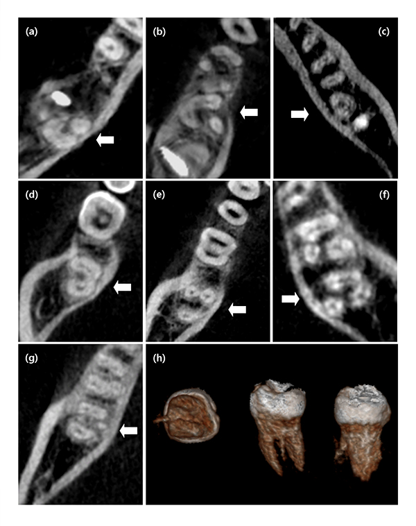
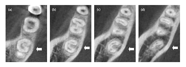
Of the 1,920 mandibular second molars, 41.15% had one root, 58.12% had two roots, and 0.72% had three roots. In the mesial roots of two-rooted molars, Vertucci's type lV (44.46%) and type Ⅱ (37.75%) canals were most frequent. The prevalence of C-shaped roots was 40.10%, and C-shaped roots in combination with additional mesiolingual or distolingual roots were found in 0.73% of molars. Interestingly, O-shaped canals were detected in 0.10% of the molars. Of the C-shaped roots, the most common configuration types were Melton’s type I (66.36%) in the coronal region and Melton’s type III (55.58%) in the apical region. The prevalence of C-shaped roots was higher in females (46.92%) than in males (32.09%) (P < 0.001) and did not differ with age (P = 0.497) or tooth position (P = 0.514). Most (82.34%) C-shaped canals were bilateral (P < 0.001).
A high prevalence of C-shaped canals and a low prevalence of three roots were observed in the mandibular second molars of a Korean population. The most frequent pattern was characterized by two separate roots with two canals in the mesial roots and one canal in the distal roots. In the mesial root of two-rooted molars, Vertucci's Type lV (two canals, two foramina) was the most commonly observed canal Type.
This is the first study to report the prevalence of rare anatomical variations, including O-shaped roots, C-shaped roots with additional ML or DL roots, and additional mid-lingual roots. These findings have not been previously described except in case reports. Evaluating CBCT scans provides a significantly better understanding of root canal anatomy, with the potential to improve the success rate of nonsurgical and surgical root canal treatments. However, CBCT should not be used routinely in all cases of endodontic treatment based on the ALARA concept.

Figure 1. Cases of mandibular second molars with root and canal variations. Arrows denote the examined teeth. (a) Mesiolingual root in combination with C-shaped root, (b) Distolingual root in combination with C-shaped root, (c) O-shaped root, (d) C-shaped root with buccally oriented groove, (e) Three roots with two mesial roots (MB, ML, and D), (f) Three roots with two distal roots (M, DB, and DL), (g) Three roots with a mid-lingual root (M, D, and Mid-lingual), (h) Three-dimensionally reconstructed images of molar with an additional mid-lingual root (apical, lingual, and distal views)

Figure 2. Cases of mandibular second molars showing change in classification at different root levels
(a) Canal orifice, C1. (b) Coronal level, C3. (c) Middle level, C2. (d) Apical level, C4.
* Related Article
S. Y. Kim, B. S. Kim, Y. Kim, Mandibular second molar root canal morphology and variants in a Korean subpopulation, International Endodontic Journal, 2016 Feb;49(2):136-44
Kim Y, Lee SJ, Woo J, Morphology of maxillary first and second molars analyzed by cone-beam computed tomography in a korean population: variations in the number of roots and canals and the incidence of fusion, Journal of Endodontics, 2012 Aug;38(8):1063-8
Kim SY, Kim BS, Woo J, Kim Y, Morphology of mandibular first molars analyzed by cone-beam computed tomography in a Korean population: variations in the number of roots and canals, Journal of Endodontics, 2013 Dec;39(12):1516-21.


