본문
Extracellular matrix-derived extracellular vesicles promote cardiomyocyte growth and electrical activity in engineered cardiac atria
by Prof. Minsuk Kim (ms@ewha.ac.kr)
Department of Pharmacology, College of Medicine
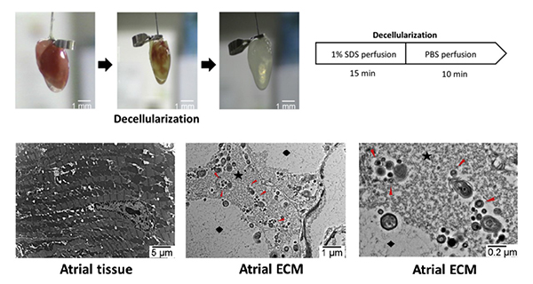
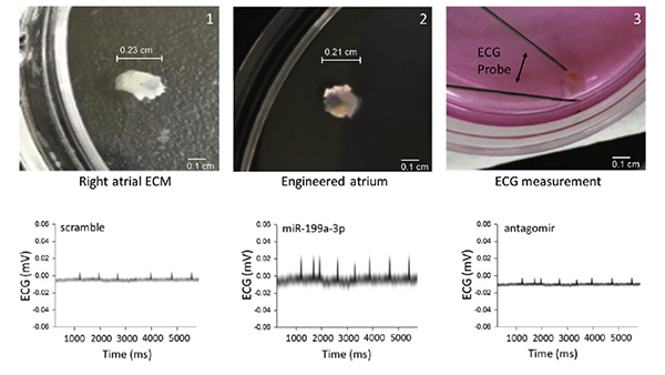
Extracellular matrix (ECM) plays a critical role in the provision of the necessary microenvironment for the proper regeneration of the cardiac tissue. However, specific mechanisms that lead to ECM-mediated cardiac regeneration are not well understood. To elucidate the potential mechanisms, we investigated ultra-structures of the cardiac ECM using electron microscopy. Intriguingly, we observed large quantities of micro-vesicles from decellularized right atria. RNA and protein analyses revealed that these contained exosomal proteins and microRNAs (miRNAs), which we referred to herein as ECM-derived extracellular vesicles (ECM-EVs). One particular miRNA from ECM-EVs, miR-199a-3p, promoted cell growth of isolated neonatal cardiomyocytes and sinus nodal cells by repressing homeodomain-only protein (HOPX) expression and increasing GATA-binding 4 (Gata4) acetylation. To determine the mechanisms, we knocked down Gata4 and showed that miR-199a-3p actions required Gata4 for cell proliferation in isolated neonatal cardiomyocytes and sinus nodal cells. To further explore the role of this miRNA, we isolated neonatal cardiac cells and recellularized into atrial ECM, referred here has engineered atria. Remarkably, miR-199a-3p mediated the enrichment of cardiomyocyte and sinus nodal cell population, and enhanced electrocardiographic signal activity of sinus nodal cells in the engineered atria. Importantly, antisense of miRNA (antagomir) against miR-199a-3p was capable of abolishing these actions of miR-199a-3p in the engineered atria. In conclusion, these results provide clear evidence of the critical role of ECM, in not only providing a scaffold for cardiac tissue growth, but also in promoting atrial electrical function through ECM-derived miR-199a-3p.

Figure 1. Ultrastructure of a decellularized heart showing interstitial matrix (star) and empty space (diamond) once occupied by cells and small size vesicles (arrows) in the atrial ECM.

Figure 2. Atrial ECMs were recellularized with neonatal cardiomyocyte, fibroblasts and sinus nodal cells. Subsequently, the engineered atria were exposed to scramble, miR-199a-3p expression plasmid or antagomir of miR-199a-3p and then ECG was determined.
* Related Article
Minae An, Kihwan Kwon, Junbeom Park, Dong-Ryeol Ryu, Jung-A. Shin, Jihee Lee Kang, Ji Ha Choi, Eun-Mi Park, Kyung Eun Lee, Minna Woo, Minsuk Kim, Extracellular matrix-derived extracellular vesicles promote cardiomyocyte growth and electrical activity in engineered cardiac atria, Biomaterials, 2017;146;49-59
Ott HC, Matthiesen TS, Goh SK, Black LD, Kren SM, Netoff TI and Taylor DA, Perfusion-decellularized matrix: using nature's platform to engineer a bioartificial heart. Nature Medicine, 2008;14:213-21.


